


Mammogram
Mammography is a specific breast imaging modality that uses low dose x-rays to detect cancer. Getting annually screened by mammography for women over the age of 40 based on the American Medical Association (AMA) and the American College of Radiology (ACR) recommendations is the best way to detect early changes even before women experience symptoms when it is most treatable. 80-90% of Breast Cancer cases can experience full recovery if detected at an early stage.
Breast Ultrasound
Ultrasound is an important modality in breast imaging and is the usual initial investigation in young or pregnant patients (usually under 30 years of age).
Ultrasound Guided Biopsy
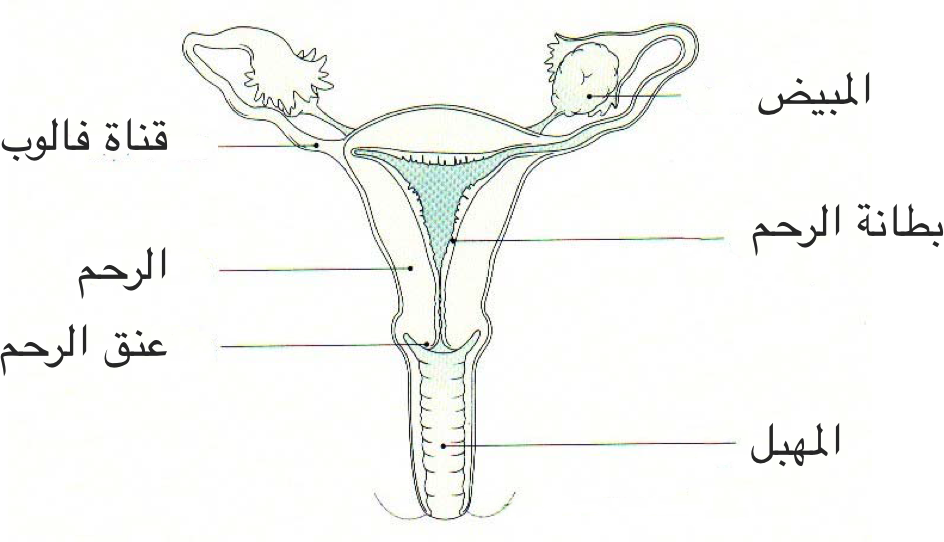
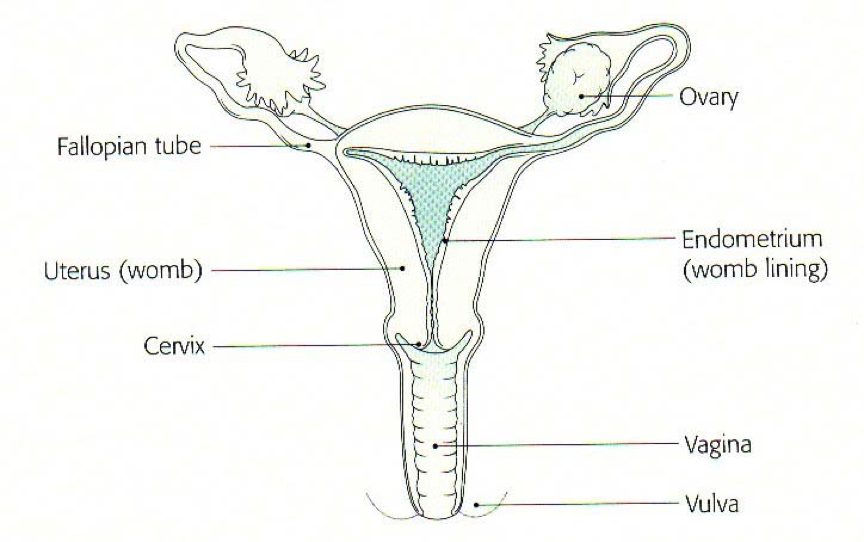
Hysteroscopy is a procedure which uses a fine telescope to examine the lining and shape of the uterus.
What are the benefits of having a Hysteroscopy?
A hysteroscopy can help to find the cause of problems relating to:
- Heavy vaginal bleeding
- Irregular periods
- Bleeding between periods
- Bleeding after sexual intercourse
- Bleeding after menopause
- Persistent discharge
- Scar tissue in the womb
- Infertility
The hysteroscope can also be used in the treatment of various intrauterine pathology such as:
- Fibroids (growths in the uterus which are not cancer)
- Polyps (blood-filled growths which are not cancer)
- Thickening of the lining of the uterus (the endometrium)
- Removal of displaced intrauterine contraceptive devices
- Removal of scar tissue.
What happens during the procedure?
A general anesthetic is used so you will be asleep and not feeling any pain. Your cervix may then be opened slightly using a smooth instrument called a dilator.
The hysteroscope (a small, fibre-optic ‘telescope’, which is attached to a small camera) is passed along your vagina and through your cervix in order to look at the inside of your uterus.
Sterile fluid is then run into your uterus to expand it to help seeing the lining of the uterus. A small sample of the lining may be removed as a biopsy depending on your condition.
If you are being treated for fibroids or polyps, an instrument called a resectoscope will be used to fully or partially remove them.
The tissue from the biopsy and the removed fibroid or polyp is then sent to the laboratory to be examined. The procedure takes between 10 and 30 minutes.
After the Procedure
When you wake up, you might feel some mild, period-like pain or cramps. Some discomfort is to be expected after the procedure for which pain relief is usually sufficient. It is important that you take your pain relief on a regular basis for the first few days. When taken regularly, the medicine is kept at a constant level in your body, so it will control your pain better. After a 1-2 days, you can gradually reduce the medicine until you do not need it any longer.